Due to the use of electrons, the images are black-and-white with grey tones depending either on the distance the electrons have to travel from the surface of the analysed sample to the detector, or on the energy of the electrons; the shorter the distance, the higher the energy, and the brighter the nuance.
Two types of images
Typically, two types of images can be collected, depending on type of electron the detector is recording:
- secondary electron (SE) images describe the topography of the analyzed surface
- backscattered electron (BSE) images contain descriptive information about differences in chemical composition
Detecting chemical elements
During the interaction of the electron beam with the sample, X-Ray beams are also generated that are recorded by the EDX detector. The energy of the recorded X-Ray beams is specific for every chemical element of the periodic table.
The instrument at the SciCult-KHM laboratory allows detection of the elements spanning form Boron to Uranium in different modes: single points measurements, area measurements, area mappings and line mappings.
Studying a broad range of materials
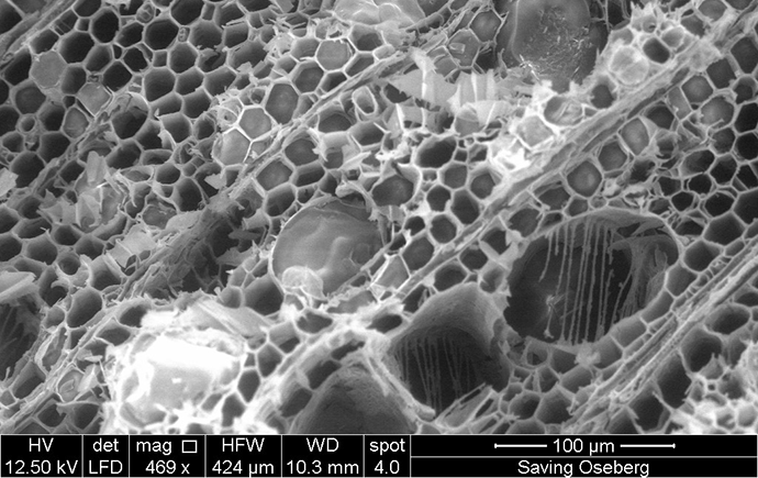
Typically, the scanning electron microscope (SEM) is used to study a very broad range of materials (from wood, leather, plastics, stone, etc.), the technology and manufacturing history of museum objects at high magnification, where fine details are revealed.
Normally, small samples (e.g. small fragments, cross-sections, threads, etc.) are placed on stubs on a sample stage, but the small object can be also accommodated by the microscope chamber.


For high and low vacuum modes
Our instrument can be used in both high and low vacuum modes.
In the cultural heritage field, the low vacuum mode is usually preferred because it allows to examine any material directly, whether it is non-conducting – such as glass, ceramics, paper, shell, charcoal, corrosion products etc. – or conducting – such as metals – without the application of a surface coating (such as carbon or gold) which is usually required for the high vacuum mode.